X-ray of the chest organs
A chest x-ray is conducted to elucidate the origins of pulmonary hypertension (PH), identify interstitial lung diseases, acquired and congenital heart defects, and assess the severity of PH.
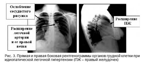
Key radiological indicators of PH include the prominence of the trunk and left branch of the pulmonary artery, shaping the second arch in the direct projection along the left contour of the heart, expansion of the lung roots, and enlargement of the right portions of the heart.
Characteristic changes are observed in 90% of cases of idiopathic pulmonary hypertension. These changes manifest as an increased transparency of the pulmonary fields at the periphery, resulting from the depletion of the pulmonary pattern. X-ray examination can reveal alterations in the lungs and heart that may contribute to the onset of pulmonary hypertension.